
when C. DIFF toxins aren’t around
The colon reabsorbs water and key electrolytes (e.g., Na⁺, Cl⁻) to maintain hydration and electrolyte balance. This allows for the production of waste in the form of stool. The gut microbiome ferments indigestible dietary fibers/polysaccharides into short-chain fatty acids like butyrate, which nourish/fuel colonocytes, help regulate immune responses, suppress inflammation, and maintain gut barrier integrity. (Mutchler, 2024)
Gut-associated lymphoid tissue (GALT) detects harmful bacteria and promotes tolerance to beneficial microbes (commensal bacteria) and immune responses in order to maintain gut homeostasis. This prevents bacterial infections via phagocytosis, antigen-presenting cell activation, and controlled cytokine release (e.g, IL-10). Secretory mucosal immunoglobulin A (IgA), produced by GALT, neutralizes pathogens without excessive inflammation. (Bemark, 2024)
The kidneys filter out waste in the form of excess sodium, potassium, and acids to maintain blood homeostasis and electrolyte balance. They additionally filter bacterial toxins and inflammatory cytokines. (Arrich, 2005) Aquaporins in the renal tubules allow reabsorption of water to prevent dehydration. The kidneys also regulate blood pressure through the renin-angiotensin system. (Suzumoto, 2023)

The Enteric Nervous System controls peristalsis and secretion via neurotransmitters like serotonin (5-HT). This system, that lines the gut wall of the small intestine, prevents constipation or excessive diarrhea by adjusting motility accordingly as well as communicates gut status to the brain via the vagus nerve. This allows for the regulation of gut movement, secretion, and pain perception. (Kulkarni, Fig. 1, right, 2018)
normal cell function
In the steady-state, intestinal bacteria are segregated from epithelial cells (IECs) by an intact mucosal layer. A well-balanced relationship is maintained between intestinal microbiota and mucosal barriers during gut homeostasis, so that gut microbes and host immune cells can mutually communicate to regulate the functions of intestinal epithelium. Commensal bacteria and pathogens can modulate the intestinal immune response to release various chemokines and cytokines, or inhibit their production, such as IL-8 and MCP-1. In a homeostasis state, pathogen recognition leads to inducing antigen-presenting cells (APCs), like macrophages, dendritic cells (DC), and neutrophils, which produce pro-inflammatory cytokines like IL-1β and IL-23. In contrast, commensals can stimulate APCs to promote anti-inflammatory cytokines, like IL-10 and regulatory T cell responses, which suppress the immune response by inhibiting cytokine production, therefore homeostasis and self-tolerance are maintained. In some cases, the interactions between commensal bacteria and the gut epithelium may lead to the discharge of TGF-β from macrophages, which triggers fibroblast proliferation (tissue remodeling). (Raeisi, 2022)
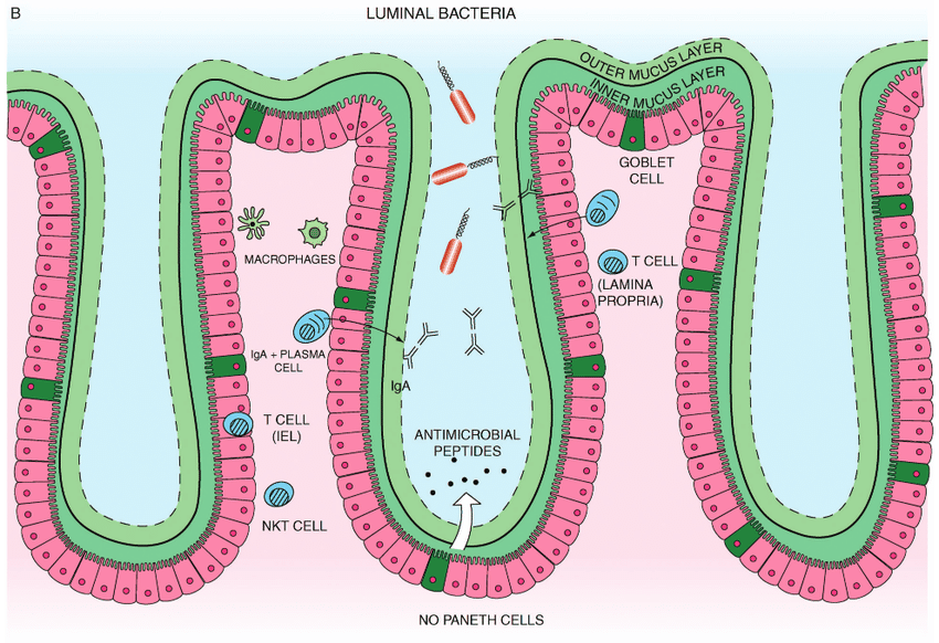
Goblet cells, seen above, produce mucins (MUC2), forming a protective mucus layer that prevents pathogens from reaching the surface of epithelial cells, therefore preventing adherence. (Mutchler, 2024) Intestinal epithelial cells, aka enterocytes line the intestinal walls and absorb vitamins, monosaccharides, minerals, ions, and amino acids for systemic energy metabolism of nutrients. They also recognize microbial ligands via pattern recognition receptors (PRRs like TLRs), and trigger appropriate immune signaling (e.g., low-level IL-8 to recruit immune cells) while maintaining barrier integrity. (Nwako, 2024) APCs (dendritic cells and macrophages) maintain balance between pro-inflammatory (IL-1β, IL-23) and anti-inflammatory responses (IL-10, TGF-β) and promote regulatory T cells for tolerance to commensals (Raeisi, 2022)
inside healthy cells
Endosomes and lysosomes in immune cells process antigens for presentation via MHC-II to helper T cells. (Nwako, 2024)
The ER is critical for folding and disulfide bond formation in proteins. It is also required for glycosylation of secreted proteins (e.g., mucins, cytokines) and calcium storage for cell signaling. It interacts with mitochondria via MAMs (mitochondria-associated membranes) for energy signaling and apoptosis regulation. (Zhang, 2020)
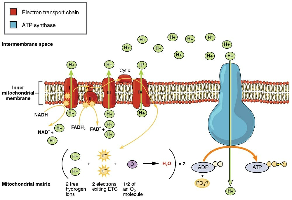
Mitochondria provide ATP via oxidative phosphorylation and the electron transport chain (pictured to the right), supporting active transport (e.g., Na⁺/K⁺ pumps in IECs) and epithelial renewal. They also generate reactive oxygen species (ROS) at low levels as signaling molecules for cell proliferation and host defense mechanisms (ROS kill microbes during infection). (Chan, 2018)
The nucleus is the site of transcription for immune-related genes in response to microbial cues. These include the NF-κB signaling pathway which is activated by microbial components via TLRs and induces transcription of cytokines like IL-8, which recruits neutrophils for infection control. As well as the PI3K-Akt signaling pathway that promotes epithelial survival and barrier maintenance; and the TGF-β pathway that promotes tolerance and suppresses inflammation, contributes to tissue remodeling and wound healing. The NF-κB Pathway is a key regulator of immune response genes. It is triggered mildly by commensals to maintain tolerance but activated strongly during infection. (Wullaert, 2011)
proper proteins
Rho-GTPases act as molecular switches that regulate actin cytoskeleton dynamics (e.g., tight junction formation and maintenance), cell polarity, endocytosis and vesicle trafficking, and barrier integrity of intestinal epithelial cells (IECs). Active (GTP-bound) forms of RhoA, Rac1, and Cdc42 promote F-actin polymerization, microvilli formation, and tight junction stability (through regulation of ZO-1 and occludin). (Arnold, 2017) The GTP-bound active state is generated by guanine exchange factors (GEFs), promoting the exchange of GDP for GTP. (Ortiz-Sanchez, Fig. 1 below, 2012)

Tight junction proteins (ZO-1, occludin, claudins) control permeability by sealing spaces between IECs, allowing nutrients in while blocking harmful pathogens, therefore preventing infection and inflammation. (Nusrat, 2001) The scaffold proteins also recognize microbial signals and trigger a mild immune response to maintain homeostasis. (Nwako, 2024) The Tight Junction Complex is composed of proteins (e.g., ZO-1, occludin, claudins), maintaining paracellular barrier integrity between epithelial cells. (Nusrat, 2001)
NOX enzymes (e.g., NOX1, NOX2) are expressed in IECs and phagocytes. They generate ROS in a regulated manner to eliminate pathogens and signal epithelial renewal and wound repair. These enzymes interact with pattern recognition receptors (PRRs) to fine-tune inflammatory responses. (Lambeth, 2004)
TLRs (e.g., TLR4) detect microbial LPS and activate MyD88-dependent NF-κB signaling which leads to controlled cytokine production. IL-10 & TGF-β are anti-inflammatory cytokines from APCs/macrophages that promote immune tolerance. (Wullaert, 2011)
MUC2 (mucin) is a glycoprotein forming the mucus layer that requires post-translational glycosylation to function. (Johansson, 2008)
SCFA transporters import SCFAs into epithelial cells to be used in energy metabolism and HDAC inhibition (epigenetic regulation). (Tan, 2024) Intermolecular bonds like hydrogen bonds and electrostatic interactions maintain the structure of tight junctions. Glycosidic bonds in mucins support mucus viscosity and resilience. Intramolecular Bonds like disulfide bridges in mucins help maintain tertiary structure. Peptide bonds in cytokines and chemokines maintain active conformation. (Tan, 2024)
Essential small molecules include O₂ which is used in mitochondrial respiration for ATP production, SCFAs (e.g., butyrate) which are used to fuel colonocytes and regulate gene expression, H₂O which is absorbed by the colon to maintain hydration, Na⁺/K⁺ which are reabsorbed to maintain electrolyte balance, and glucose and amino acids which are absorbed and used for systemic metabolism and repair. (Tan, 2024)
references
- Health. Mutchler “Why Your Large Intestine Is Such a Big Deal.” Accessed March 31, 2025. https://www.health.com/large-intestine-8706814.
- Nwako, Jennifer G., and Heather A. McCauley. “Enteroendocrine Cells Regulate Intestinal Homeostasis and Epithelial Function.” Molecular and Cellular Endocrinology 593 (November 1, 2024): 112339. https://doi.org/10.1016/j.mce.2024.112339.
- Bemark “Gut-Associated Lymphoid Tissue: A Microbiota-Driven Hub of B Cell Immunity – PMC.” Accessed March 31, 2025. https://pmc.ncbi.nlm.nih.gov/articles/PMC11227984/.
- Suzumoto, Yoko, Laura Zucaro, Anna Iervolino, and Giovambattista Capasso. “Kidney and Blood Pressure Regulation—Latest Evidence for Molecular Mechanisms.” Clinical Kidney Journal 16, no. 6 (June 1, 2023): 952–64. https://doi.org/10.1093/ckj/sfad015.
- Kulkarni “Advances in Enteric Neurobiology: The ‘Brain’ in the Gut in Health and Disease | Journal of Neuroscience.” Accessed March 31, 2025. https://www.jneurosci.org/content/38/44/9346.
- Voth, Daniel E., and Jimmy D. Ballard. “Clostridium Difficile Toxins: Mechanism of Action and Role in Disease.” Clinical Microbiology Reviews 18, no. 2 (April 2005): 247–63. https://doi.org/10.1128/CMR.18.2.247-263.2005.
- Wullaert, Andy, Marion C. Bonnet, and Manolis Pasparakis. “NF-κB in the Regulation of Epithelial Homeostasis and Inflammation.” Cell Research 21, no. 1 (January 2011): 146–58. https://doi.org/10.1038/cr.2010.175.
- Zhang, Irina X., Malini Raghavan, and Leslie S. Satin. “The Endoplasmic Reticulum and Calcium Homeostasis in Pancreatic Beta Cells.” Endocrinology 161, no. 2 (February 1, 2020): bqz028. https://doi.org/10.1210/endocr/bqz028.
- Raeisi, Hamideh, Masoumeh Azimirad, Ali Nabavi-Rad, Hamid Asadzadeh Aghdaei, Abbas Yadegar, and Mohammad Reza Zali. “Application of Recombinant Antibodies for Treatment of Clostridioides Difficile Infection: Current Status and Future Perspective.” Frontiers in Immunology 13 (August 23, 2022). https://doi.org/10.3389/fimmu.2022.972930.
- Arnold, Torey R., Rachel E. Stephenson, and Ann L. Miller. “Rho GTPases and Actomyosin: Partners in Regulating Epithelial Cell-Cell Junction Structure and Function.” Experimental Cell Research 358, no. 1 (September 1, 2017): 20–30. https://doi.org/10.1016/j.yexcr.2017.03.053.
- Tan, Aolei, and Xiaoxiao Ma. “Exploring the Functional Roles of Small-Molecule Metabolites in Disease Research: Recent Advancements in Metabolomics.” Chinese Chemical Letters 35, no. 8 (August 1, 2024): 109276. https://doi.org/10.1016/j.cclet.2023.109276.
- Nusrat, A., C. von Eichel-Streiber, J. R. Turner, P. Verkade, J. L. Madara, and C. A. Parkos. “Clostridium Difficile Toxins Disrupt Epithelial Barrier Function by Altering Membrane Microdomain Localization of Tight Junction Proteins.” Infection and Immunity 69, no. 3 (March 2001): 1329–36. https://doi.org/10.1128/IAI.69.3.1329-1336.2001.
- Lambeth, J. David. “NOX Enzymes and the Biology of Reactive Oxygen.” Nature Reviews. Immunology 4, no. 3 (March 2004): 181–89. https://doi.org/10.1038/nri1312.
- Johansson “The Inner of the Two Muc2 Mucin-Dependent Mucus Layers in Colon Is Devoid of Bacteria – PubMed.” Accessed April 20, 2025. https://pubmed.ncbi.nlm.nih.gov/18806221/.
- Arrich, Jasmin, Gottfried-H. Sodeck, Gurkan Sengolge, Christoforos Konnaris, Marcus Mullner, Anton-N. Laggner, and Hans Domanovits. “Clostridium Difficile Causing Acute Renal Failure: Case Presentation and Review.” World Journal of Gastroenterology 11, no. 8 (February 28, 2005): 1245–47. https://doi.org/10.3748/wjg.v11.i8.1245.
- Unsplash. “Body Anatomy Pictures | Download Free Images on Unsplash.” Accessed April 21, 2025. https://unsplash.com/s/photos/body-anatomy.
- Soveral, Lucas F., Gabriela G. Korczaguin, Pedro S. Schmidt, Isabel S. Nunes, Camilo Fernandes, and Carlos R. Zárate-Bladés. “Immunological Mechanisms of Fecal Microbiota Transplantation in Recurrent Clostridioides Difficile Infection.” World Journal of Gastroenterology 28, no. 33 (September 7, 2022): 4762–72. https://doi.org/10.3748/wjg.v28.i33.4762.
- Editors, B. D. “Electron Transport Chain and Oxidative Phosphorylation.” Biology Dictionary (blog), August 22, 2018. https://biologydictionary.net/electron-transport-chain-and-oxidative-phosphorylation/.
- AhmadkruwHickman. “Which Cells in the Small Intestine’s Mucosa Secrete Mucus,” April 17, 2022. https://ahmadkruwhickman.blogspot.com/2022/04/which-cells-in-small-intestines-mucosa.html.
- Ortiz-Sanchez “Identification of Potential Small Molecule Binding Pockets on Rho Family GTPases | PLOS One.” https://journals.plos.org/plosone/article?id=10.1371/journal.pone.0040809.