The species name “difficile” is derived from the Latin word for “difficult.”
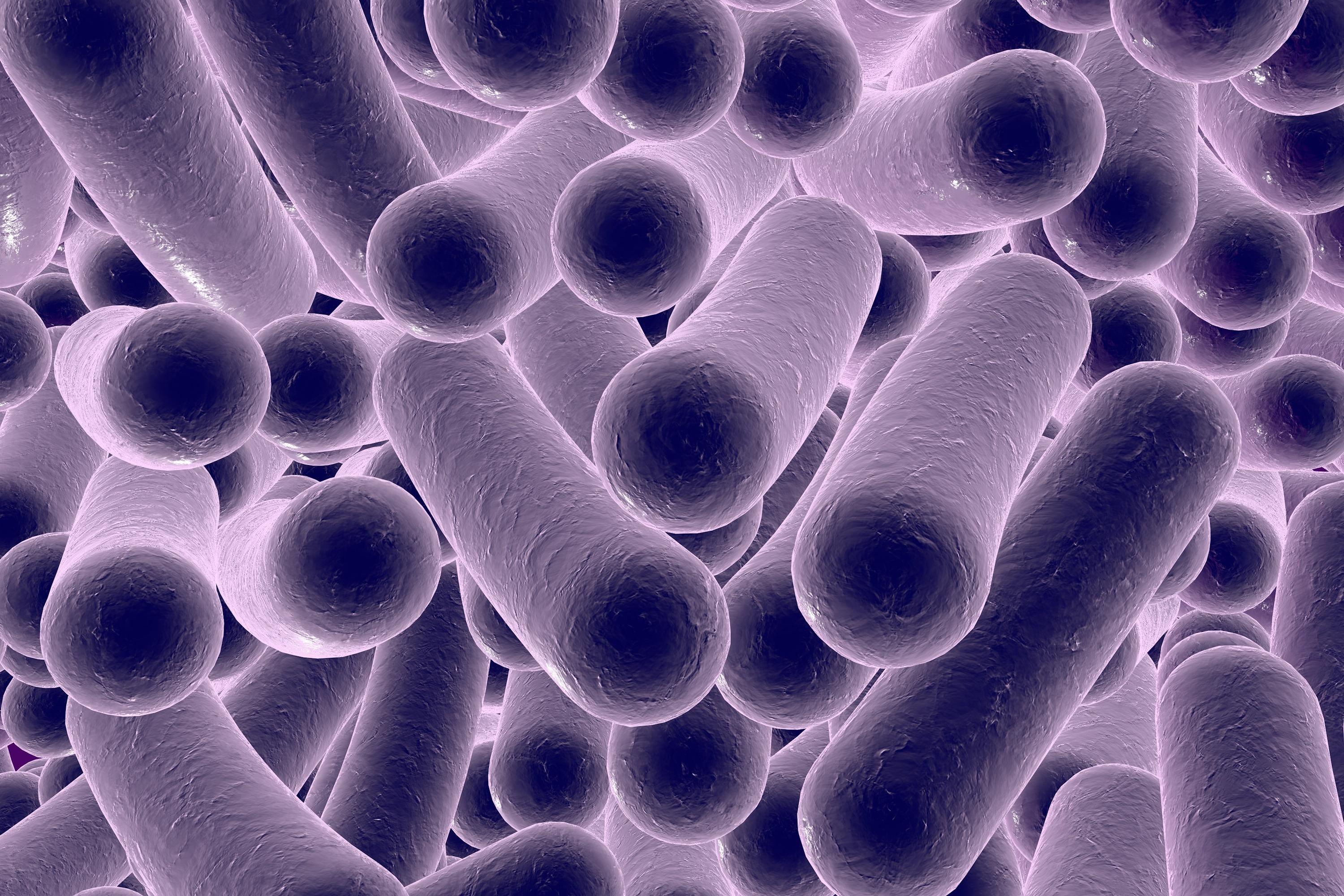
This name was given because C. difficile was notoriously hard to isolate and grow in lab cultures when it was first discovered in 1935 by Hall and O’Toole. (Hall, 1935) Originally classified under Clostridium, it was later reclassified as Clostridioides based on a genetic analysis. The name Clostridium comes from the Greek klōstēr, meaning “spindle,” referencing the rod-shaped appearance of these bacteria. Studies using 16S rRNA sequencing and whole-genome analysis showed that C. difficile was genetically distinct from the core Clostridium species. It had significant differences in its evolutionary lineage compared to other members of the genus. Additionally, Clostridium was a broad genus containing many unrelated species. Scientists determined that C. difficile was more closely related to other bacteria outside of Clostridium, leading to its placement in a new genus, Clostridioides. The reclassification, proposed in 2016 by Lawson et al., aimed to create a more accurate and evolutionarily meaningful bacterial taxonomy. So, Clostridioides difficile essentially means a “difficult-to-grow, spindle-shaped bacterium.” (The Lancet, 2019)
the first findings
Ivan C. Hall and Elizabeth O’Toole were the first scientists to report C.diff in 1935. It was located during a bacterial study of infant stool during the first 10 days of life. They regarded this finding as inevitable from the infant coming into contact with the vaginal flora of the mother, her nipples, and from other sources of bacteria. (Hall, 1935)
Almost 30 years later, in 1962, Louis Ds. Smith and Elizabeth O. King isolated C. diff strains from infections like gas gangrene, abscesses and fluid. These strains were regarded as non-pathogenic. (Smith,1962) It wasn’t until 1978 that C.diff strains were isolated from patients suffering from pseudomembranous colitis due to antibiotic use and determined to be causing cytotoxicity by John G. Bartlett, Te-Wen Chang, Marc Gurwith, Sherwood L. Gorbach, and Andrew B. Onderdonk. (Bartlett,1978)

Sherwood Gorbach and Te-Wen Chang of the landmark 1978 paper honored alongside their Tufts colleague for their distinguished achievements in the field of medicine and infectious disease. Harris Berman, dean of the medical school, presented them with Dean’s Medals in 2015.
In his prefatory remarks, Berman was quick to observe that the evening’s award recipients were “people who made a significant difference not just at Tufts, but in the world.” (TuftsNow, 2015)

methods for isolation
In their 1978 landmark study, Bartlett et al. identified the cytotoxic effects of Clostridium difficile using cell culture assays and toxin neutralization tests. In order to do this, they collected stool samples from patients suffering from antibiotic-associated pseudomembranous colitis and isolated the C.diff strains present in their stool. They cultured the bacterial strains in anaerobic conditions and obtained culture filtrates (liquid media containing bacterial secretions, including toxins). They applied the filtrates to monolayers of cultured mammalian cells. The cells exhibited characteristic cytopathic effects, such as rounding, shrinkage, and detachment, indicating toxin-induced damage. To confirm that the observed cell damage was caused by a toxin and not some other factor, they added Clostridium sordellii antitoxin to the filtrates before exposing them to the cells. (Chang,1978) The antitoxin neutralized the cytotoxic effects, proving that the damage was due to a toxin produced by the bacterial strains. This study was crucial in establishing C. diff as the causative agent of antibiotic-associated pseudomembranous colitis, linking its cytotoxic toxins to the disease pathology. (Bartlett,1978)
They noticed Clostridium difficile as a disease-causing organism because of the severe gastrointestinal symptoms seen in patients following antibiotic treatment. Before the discovery of C. diff, pseudomembranous colitis was recognized as a complication of antibiotic use, but the cause was unknown. (Bartlett,1978)
Patients with a C. diff infection typically exhibit the following symptoms: (CDC, 2024)
Profuse, Watery Diarrhea – The most common symptom, often severe and frequent (sometimes more than 10 times a day).
Abdominal Pain and Cramping – Due to inflammation and toxin-induced damage to the intestinal lining. Patients often experience tenderness and bloating.
Fever – A sign of systemic inflammation due to the bacterial infection. Pseudomembranous Colitis – Characterized by yellowish-white plaques (pseudomembranes) on the colon’s inner surface, seen via colonoscopy. These are formed from dead cells, mucus, and fibrin.
Leukocytosis– Blood tests often show an abnormally high number of white blood cells, indicating an immune response.
Toxic Megacolon and Sepsis –In extreme cases, the colon becomes massively distended and stops functioning, which can be life-threatening. If toxins enter the bloodstream, they can lead to sepsis and organ failure.
why people actually cared

pseudomembranous-colitis-light-micrograph
-medimagescience-photo-library.html.
Doctors observed that patients who had recently taken broad-spectrum antibiotics (like clindamycin or ampicillin) developed sudden, severe diarrhea and the priorly listed symptoms. Colonoscopies revealed pseudomembranes, a striking and unusual sign of colitis. The scientists completed stool studies on these patients in order to determine the cause of these pseudomembranes, leading them to isolate abnormally present C. diff toxins as the causative agent. This was typically done using the Enzyme Immunoassay method which can detect substances produced by bacteria as well as the cell culture cytotoxicity neutralization assay method which examines the cytopathic effects of these toxins on cultured cells. Today, scientists use a more sensitive method, polymerase chain reaction, to detect toxin genes directly. (O’Connor, 2001) Based on their findings, C. diff became recognized as a major cause of antibiotic-associated diarrhea and colitis, leading to further research into its toxins and treatments. (Lance George, 1978)
The studies on Clostridium difficiles’ toxicity occurred because doctors and researchers were trying to understand a mysterious and severe form of antibiotic-associated diarrhea known as pseudomembranous colitis (pictured to the right). In the 1960s and 1970s, doctors noticed that many patients who took broad-spectrum antibiotics developed severe, persistent diarrhea. Some cases were so serious that they led to dehydration, sepsis, and even death. (Bartlett, 1978) C.diff was, and still is, most common amongst elderly patients, especially those who have spent extended periods of time in a hospital setting, where exposure and antibiotic use is high. As elderly patients are already more susceptible to disease, it is easily contracted by this population, leading to abnormally high numbers of deaths from C.diff infection due to the fact that it is most common in those already suffering from a secondary disease or simply age progression. Scientists and doctors wanted to find a way to ease the suffering of their patients, lessen contagious exposure in hospitals, and determine why antibiotic use causes C.diff infection in some but not others. (CDC, 2024)
references
- HALL, IVAN C., and ELIZABETH O’TOOLE. “INTESTINAL FLORA IN NEW-BORN INFANTS: WITH A DESCRIPTION OF A NEW PATHOGENIC ANAEROBE, BACILLUS DIFFICILIS.” American Journal of Diseases of Children 49, no. 2 (February 1, 1935): 390–402. https://doi.org/10.1001/archpedi.1935.01970020105010.
- Diseases, The Lancet Infectious. “C Difficile—a Rose by Any Other Name….” The Lancet Infectious Diseases 19, no. 5 (May 1, 2019): 449. https://doi.org/10.1016/S1473-3099(19)30177-X.
- Smith, Louis Ds., and Elizabeth O. King. “OCCURRENCE OF CLOSTRIDIUM DIFFICILE IN INFECTIONS OF MAN.” Journal of Bacteriology 84, no. 1 (July 1962): 65–67. https://doi.org/10.1128/jb.84.1.65-67.1962
- Bartlett, John G., Te Wen Chang, Marc Gurwith, Sherwood L. Gorbach, and Andrew B. Onderdonk. “Antibiotic-Associated Pseudomembranous Colitis Due to Toxin-Producing Clostridia.” New England Journal of Medicine 298, no. 10 (March 9, 1978): 531–34. https://doi.org/10.1056/NEJM197803092981003.
- Chang, T W, S L Gorbach, and J B Bartlett. “Neutralization of Clostridium Difficile Toxin by Clostridium Sordellii Antitoxins.” Infection and Immunity 22, no. 2 (November 1978): 418–22. https://doi.org/10.1128/iai.22.2.418-422.1978
- O’Connor DHynes P, Cormican MCollins E, Corbett-Feeney G, Cassidy M.2001.Evaluation of Methods for Detection of Toxins in Specimens of Feces Submitted for Diagnosis of Clostridium difficile- Associated Diarrhea. J Clin Microbiol39:.https://doi.org/10.1128/jcm.39.8.2846-2849.2001
- Lance George, W., EllieJ. C. Goldstein, VeraL. Sutter, ShellyL. Ludwig, and SydneyM. Finegold. “ÆTIOLOGY OF ANTIMICROBIAL-AGENT-ASSOCIATED COLITIS.” The Lancet, Originally published as Volume 1, Issue 8068, 311, no. 8068 (April 15, 1978): 802–3. https://doi.org/10.1016/S0140-6736(78)93001-5.
- “Honoring Tufts Pioneers in Medicine | Tufts Now,” November 13, 2015. https://now.tufts.edu/2015/11/13/honoring-tufts-pioneers-medicine.
- CDC. “About C. Diff.” <em>C. diff </em>(<em>Clostridioides difficile</em>), December 19, 2024. https://www.cdc.gov/c-diff/about/index.html.
- “Clostridiodes Difficile (C. Diff): Symptoms & Treatment.” Accessed April 21, 2025. https://www.health.com/c-diff-overview-7485174.
- Pixels. “Pseudomembranous Colitis Light Micrograph by Science Photo Library.” Accessed April 21, 2025. https://pixels.com/featured/pseudomembranous-colitis-light-micrograph-medimagescience-photo-library.html.
- “C. Diff Infections.” Text. National Library of Medicine. Accessed April 22, 2025. https://medlineplus.gov/cdiffinfections.html.