The primary target

The colon is the primary site of infection and pathology for C.difficile. Toxins A and B produced by C. diff cause damage to the intestinal epithelial cells through the inactivation of Rho GTPases, leading to cytoskeletal breakdown and the loss of cell-cell junction integrity. This also increases intestinal permeability, allowing fluids, toxins, and immune cells to leak into the gut lumen, which results in diarrhea and malabsorption. The action of toxins triggers the activation of the NF-κB pathway. Unregulated NF-κB activation causes excessive cytokine release, neutrophil influx, and hyperinflammation. (Just, 1995) This delays patient recovery and results in inflammatory cell infiltration into the colon, contributing to ulcerations and the formation of pseudomembranes. (Bartlett, 1978) Abdominal pain and cramping stem from this inflammation, ulceration, and swelling of the colon. The ongoing inflammatory process, combined with cell death, leads to tissue necrosis, further worsening inflammation and damage. In severe cases, this can progress to toxic megacolon—a massive dilation of the colon that may impair motility and function. Figure 1 to the right shows a compound tomography (CT) scan of patient with C.diff associated colitis showing diffuse thickening along the entire colon. (Madan, Fig. 1 to the left {above on mobile}, 2012) The colon’s ability to absorb water and electrolytes is additionally compromised due to inflammatory damage to microvilli and the loss of intestinal barrier function. This leads to diarrhea and electrolyte imbalances, especially potassium depletion, which can be severe in chronic cases. Bloody stools and mucus can be seen in severe cases, correlating with the destruction of mucosal tissue. (Lance George, 1978)
Casulaties of war
Toxins can also affect the enteric nervous system, disrupting normal gut motility and contributing to gut stasis (slowed bowel movement). This can worsen constipation or abdominal distention, particularly in the case of toxic megacolon, where the colon becomes massively dilated and loses its ability to contract. Delayed gastric emptying and intestinal paralysis may exacerbate symptoms. (Lance George, 1978) While the primary site of infection is the colon, the systemic effects of inflammation and immune activation can affect other tissues and organs. (Madan, 2012) For example, high inflammatory cytokine levels overwhelm renal clearance. Severe dehydration, caused by persistent diarrhea and fluid loss, can also contribute to renal dysfunction. If the infection causes significant fluid and electrolyte loss (especially potassium), acute kidney injury can develop. Symptoms like fatigue, muscle weakness, and confusion can arise due to this electrolyte imbalance. (Arrich, 2005)
The least understood part of the body, the microbiome. Essential to all, deadly to some.
Most people think of bacteria as harmful germs that make us sick, but that’s only part of the story. In reality, our bodies—especially our gut—are home to trillions of bacteria that play essential roles in digestion, immunity, and overall health. This collection of microbes, known as the microbiome, thrives on balance. While we often hear about “good” and “bad” bacteria, it’s not just about eliminating the bad—it’s about maintaining the right mix. Even some bacteria we consider harmful can be harmless or even helpful in small amounts. When this balance is disrupted, it can lead to health problems, including infections and chronic conditions. So, not all bacteria are villains—many are actually vital allies.
Some are luckier than others
C. difficile is a spore-forming, toxin-producing bacterium that can colonize the human gut microbiome without necessarily causing disease. A healthy, diverse gut microbiota plays a critical role in resisting C. difficile colonization. This concept, known as “colonization resistance,” involves microbial competition for nutrients and space, as well as the production of antimicrobial substances. Disruption of this microbiota—often due to antibiotic use—removes this protective barrier, allowing C. difficile to proliferate and produce toxins. Some individuals, particularly healthy adults and infants can harbor C. difficile without showing symptoms. In these cases, the host immune system may effectively neutralize the bacterial toxins, preventing disease. For example, the presence of antitoxin antibodies, particularly against toxin A, has been associated with protection from symptomatic infection. The pathogenicity of C. difficile is primarily due to its toxins (TcdA and TcdB). However, not all strains produce toxins, and some colonizing strains may be non-toxigenic. Individuals infected with these non-toxigenic strains typically do not develop disease. The risk of developing an actual infection increases in clinical settings where patients are exposed to antibiotics, are immunocompromised, or have other risk factors like recent surgery or prolonged hospital stays. The imbalance between mucosal barriers and gut microbes is promoted by the dysfunction of mucosal barriers, including decreased production of mucin that causes intestinal inflammation. A combination of genetic and environmental factors, especially antibiotic administration, leads to gut microbiota dysbiosis, and thereby enrichment of pathobionts and susceptibility to C. difficile may occur. Outside of these conditions, even if C. difficile spores are present, infection is less likely to occur. (Smits, 2016) The application of monoclonal antibodies (mAbs) developed for targeting toxins of C. difficile has been found to help modulate direct damage to the colonic epithelium caused by toxins and restore the homeostatic immune responses and ameliorates inflammation. (Raeisi, Fig. below, 2022)

toxin Invasion
Intestinal epithelial cells (aka goblet cells) line the colonic mucosa and are the primary target of C. diff toxins. The Combined Repetitive Oligopeptides domain of C.diff toxins allows them to bind to carbohydrate receptors on host cells, aiding in their endocytosis. The V-ATPases of these endosomes decrease cellular pH which causes a conformational change to the toxin delivery domain and leads to pore formation in the endosomal membrane. This allows for the translocation of the glucosyltransferase and autoprotease domains of the toxins into the cytosol. (Smits, 2016, Fig 5) Toxin A (TcdA) and Toxin B (TcdB), are glucosyltransferases produced by C.diff that can inactivate Rho GTPases via their glucosyltransferase domain. This inactivation disrupts the actin cytoskeleton, leading to cell rounding, loss of cell junctions, and increased permeability. (KEGG, 2025) This causes intestinal barrier dysfunction, allowing leakage of fluids and immune cells into the gut, contributing to inflammation, abdominal pain, and diarrhea. Recurrent, watery diarrhea results from this increased permeability as well as ion imbalance and loss of microvilli.
the hosts failed attempt to fight back
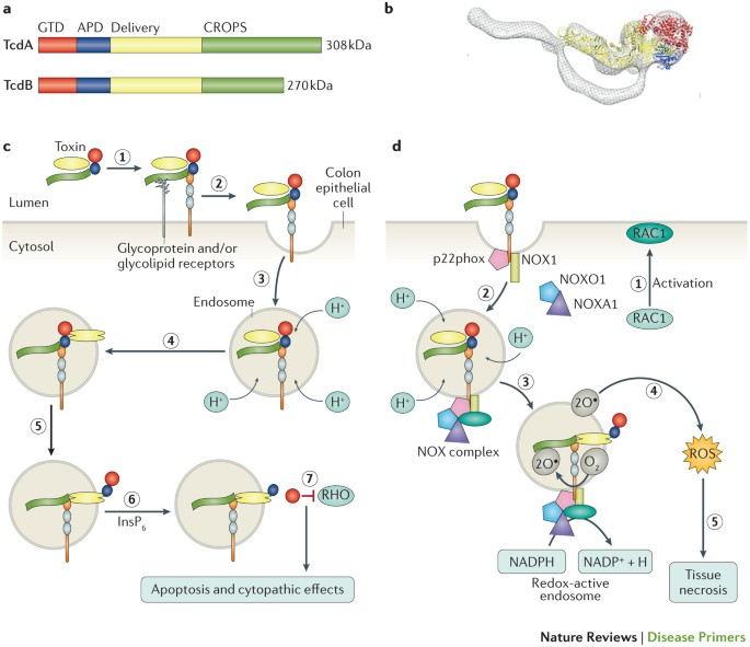
High concentrations of C.diff Toxin B leads to RAS-related C3 botulinum toxin substrate 1 (RAC1) activation and complex formation between p22phox (also known as cytochrome b-245 light chain), NADPH oxidase 1 (NOX1), NADPH oxidase activator 1 (NOXA1), NADPH oxidase organizer 1 (NOXO1) and RAC1 to make the NADPH oxidase (NOX) complex on endosome membranes. (part d of Fig. 5 to the right) This complex produces superoxide by transferring an electron from NADPH to molecular oxygen. This leads to the generation of reactive oxygen species that lead to intestinal epithelial cell death and therefore the loss of tight junction integrity. This accumulation also allows for the formation of ulcers and lesions, which are characteristic of pseudomembranous colitis. (Smits, Fig 5, to the left {above on mobile}, 2016) This ulceration and mucosal erosion can cause bloody stools in severe cases of infection. Additionally, this loss of goblet cells decreases mucus production, impairing protection against bacteria and worsening inflammation in the host. (Jank, 2007) Dysregulated NOX activation also alters redox-sensitive signaling (e.g., NF-κB) and recruits neutrophils that release further reactive oxygen species, worsening inflammation and epithelial injury.
Immune cells are activated in response to the toxin-induced damage, leading to inflammation and colonic injury. Toxin B causes a dose dependent influx in neutrophils that can be mediated by macrophage-derived TNF-α (tumor necrosis factor alpha) and lipoxygenase products. (Souza, 1997) Neutrophils, a type of white blood cell, infiltrate the colonic mucosa due to an IL-8 release, which contributes to pseudomembrane formation. (Kelly, 1994) This infiltration leads to colonic swelling and abdominal pain. These immune cells secrete cytokines as an immune response, hoping they will regulate the immune system and inflammation. But, fever and systemic inflammation seen in patients can result from excessive cytokine production, suggesting that there is stress on the system. (Kelly, 2008)
Fibroblasts in the colon typically help maintain tissue structure but become dysregulated in C.diff infection states. Toxin-induced injury activates fibroblasts, promoting collagen deposition and contributing to fibrosis. Chronic inflammation can lead to scarring and impair colonic motility. In recurrent infection cases, strictures, or the narrowing of the colon, has been seen. (Piccolo, 2016)
Mechanisms of cellular damage
The Enteric nervous system controls gut motility and secretion, but C. diff toxins can disrupt neuronal signaling. Toxin A interferes with submucosal and myenteric neurons, disrupting muscle contractions, leading to paralytic ileus or toxic megacolon. This also explains severe bloating and distention. Toxin A also disrupts the normal secretion of neurotransmitters, further impairing gut function. (Voth, 2005)
C. diff Toxin A and B glucosylate Rho-family GTPases which are crucial for regulating actin polymerization, cell shape, signaling, adhesion, and migration. This glucose modification disrupts actin polymerization, leading to cell rounding, loss of adhesion, increased permeability, and prevents cytoskeletal remodeling. This loss of barrier function, aka leaky gut, leads to diarrhea. This works through the disruption of normal fluid and electrolyte regulation due to the damage accumulated to intestinal microvilli, which enhance absorption and surface area. This disruption allows these fluids to leak from the bloodstream into the intestinal lumen and cause liquidy stools. In severe cases, leaky gut can cause sepsis. (Jank, 2007)

damage to the powerhouse of the cell????? That cant be good
TcdA and TcdB induce mitochondrial dysfunction which increases the concentration of reactive oxygen species as a byproduct. This induces oxidative damage and triggers mitochondrial permeability, leading to apoptosis and therefore inflammation. (Fiorentini, 1998) Additionally, C.diff infection allows for activation of Bax/Bak (B-cell lymphoma 2 proteins) by a BH3-only protein. This increases mitochondrial outer membrane permeability and releases cytochrome C which triggers caspase activation, leading to cell death. (Voth, 2005)
C. diff infection also indirectly triggers Endoplasmic Reticulum stress due to misfolded protein accumulation in the ER and inflammation. This is a glucosyltransferase-independent induction of stress by the toxins. This stress activates protein kinase R-like ER kinase (PERK) as part of the unfolded protein response. This impaired protein trafficking leads to abnormal cytokine secretion and increases eIF2α (eukaryotic translation initiation factor 2 alpha) signaling through phosphorylation. ElF2a regulates protein synthesis, and its phosphorylation leads to the upregulation of genes involved in protein folding and repair. This additionally leads to a decrease in protein synthesis. Calcium leakage due to ER stress causes mitochondrial overload and apoptosis. (Sun, 2014) Zonula occludens-1 (ZO-1) and occludin (membrane proteins) are also disrupted due to Rho GTPase inactivation, which weakens the epithelial barrier. This increased paracellular permeability allows for bacterial translocation and exacerbates the chance of recurrent infection. Membrane protein monolayers, control and exposed to C.diff toxin B, localized by immunofluorescence labeling and confocal microscopy, show destruction to cell integrity via the displacement of proteins from the lateral membrane for all toxin exposed cells except E-Cadherin. (Nusrat, Fig 3 to the right {above for mobile}, 2001)
references
- Lance George, W., EllieJ. C. Goldstein, VeraL. Sutter, ShellyL. Ludwig, and SydneyM. Finegold. “ÆTIOLOGY OF ANTIMICROBIAL-AGENT-ASSOCIATED COLITIS.” The Lancet, Originally published as Volume 1, Issue 8068, 311, no. 8068 (April 15, 1978): 802–3. https://doi.org/10.1016/S0140-6736(78)93001-5.
- Just, I., J. Selzer, M. Wilm, C. von Eichel-Streiber, M. Mann, and K. Aktories. “Glucosylation of Rho Proteins by Clostridium Difficile Toxin B.” Nature 375, no. 6531 (June 8, 1995): 500–503. https://doi.org/10.1038/375500a0.
- Nusrat, A., C. von Eichel-Streiber, J. R. Turner, P. Verkade, J. L. Madara, and C. A. Parkos. “Clostridium Difficile Toxins Disrupt Epithelial Barrier Function by Altering Membrane Microdomain Localization of Tight Junction Proteins.” Infection and Immunity 69, no. 3 (March 2001): 1329–36. https://doi.org/10.1128/IAI.69.3.1329-1336.2001.
- Kelly, Ciarán P., and J. Thomas LaMont. “Clostridium Difficile–More Difficult than Ever.” The New England Journal of Medicine 359, no. 18 (October 30, 2008): 1932–40. https://doi.org/10.1056/NEJMra0707500.
- Piccolo, Francesco, Anna Sze Tai, Hooi Ee, Siobhain Mulrennan, Scott Bell, and Gerard Ryan. “Clostridium Difficile Infection in Cystic Fibrosis: An Uncommon but Life‐threatening Complication.” Respirology Case Reports 5, no. 1 (December 7, 2016): e00204. https://doi.org/10.1002/rcr2.204.
- Bartlett, John G., Te Wen Chang, Marc Gurwith, Sherwood L. Gorbach, and Andrew B. Onderdonk. “Antibiotic-Associated Pseudomembranous Colitis Due to Toxin-Producing Clostridia.” New England Journal of Medicine 298, no. 10 (March 9, 1978): 531–34. https://doi.org/10.1056/NEJM197803092981003.
- Madan, Rajat, and William A. Petri. “Immune Responses to Clostridium Difficile Infection.” Trends in Molecular Medicine 18, no. 11 (November 2012): 658–66. https://doi.org/10.1016/j.molmed.2012.09.005.
- Arrich, Jasmin, Gottfried-H. Sodeck, Gurkan Sengolge, Christoforos Konnaris, Marcus Mullner, Anton-N. Laggner, and Hans Domanovits. “Clostridium Difficile Causing Acute Renal Failure: Case Presentation and Review.” World Journal of Gastroenterology 11, no. 8 (February 28, 2005): 1245–47. https://doi.org/10.3748/wjg.v11.i8.1245.
- Jank, Thomas, Torsten Giesemann, and Klaus Aktories. “Rho-Glucosylating Clostridium Difficile Toxins A and B: New Insights into Structure and Function.” Glycobiology 17, no. 4 (April 2007): 15R-22R. https://doi.org/10.1093/glycob/cwm004.
- Smits, Wiep Klaas, Dena Lyras, D. Borden Lacy, Mark H. Wilcox, and Ed J. Kuijper. “Clostridium Difficile Infection.” Nature Reviews. Disease Primers 2 (April 7, 2016): 16020. https://doi.org/10.1038/nrdp.2016.20.
- “KEGG PATHWAY: Regulation of Actin Cytoskeleton – Homo Sapiens (Human).” Accessed March 31, 2025. https://www.genome.jp/pathway/hsa04810.
- Kelly, C. P., S. Becker, J. K. Linevsky, M. A. Joshi, J. C. O’Keane, B. F. Dickey, J. T. LaMont, and C. Pothoulakis. “Neutrophil Recruitment in Clostridium Difficile Toxin A Enteritis in the Rabbit.” The Journal of Clinical Investigation 93, no. 3 (March 1994): 1257–65. https://doi.org/10.1172/JCI117080.
- Voth, Daniel E., and Jimmy D. Ballard. “Clostridium Difficile Toxins: Mechanism of Action and Role in Disease.” Clinical Microbiology Reviews 18, no. 2 (April 2005): 247–63. https://doi.org/10.1128/CMR.18.2.247-263.2005.
- Fiorentini, C., A. Fabbri, L. Falzano, A. Fattorossi, P. Matarrese, R. Rivabene, and G. Donelli. “Clostridium Difficile Toxin B Induces Apoptosis in Intestinal Cultured Cells.” Infection and Immunity 66, no. 6 (June 1998): 2660–65. https://doi.org/10.1128/IAI.66.6.2660-2665.1998.
- Sun, Chunli, Haiying Wang, Shuyi Chen, Zhendong Li, Shan Li, and Jufang Wang. “Recombinant Clostridium Difficile Toxin B Induces Endoplasmic Reticulum Stress in Mouse Colonal Carcinoma Cells.” Acta Biochimica Et Biophysica Sinica 46, no. 11 (November 2014): 973–81. https://doi.org/10.1093/abbs/gmu091.
- Souza, M H, A A Melo-Filho, M F Rocha, D M Lyerly, F Q Cunha, A A Lima, and R A Ribeiro. “The Involvement of Macrophage-Derived Tumour Necrosis Factor and Lipoxygenase Products on the Neutrophil Recruitment Induced by Clostridium Difficile Toxin B.” Immunology 91, no. 2 (June 1997): 281–88. https://doi.org/10.1046/j.1365-2567.1997.00243.x
- Raeisi, Hamideh, Masoumeh Azimirad, Ali Nabavi-Rad, Hamid Asadzadeh Aghdaei, Abbas Yadegar, and Mohammad Reza Zali. “Application of Recombinant Antibodies for Treatment of Clostridioides Difficile Infection: Current Status and Future Perspective.” Frontiers in Immunology 13 (August 23, 2022). https://doi.org/10.3389/fimmu.2022.972930.