The real enemy: sugar
C. diff Toxin A (TcdA) and Toxin B (TcdB) belong to the large clostridial toxins family and are glucosyltransferases that modify host Rho family GTPases, such as RhoA, Rac1, and Cdc42. The toxins glucosylate Rho family GTPases by catalyzing the covalent attachment of glucose molecules to key threonine residues within the GTPases. An O-glycosidic bond between UDP-glucose and the hydroxyl group of threonine in RhoA, Rac1, or Cdc42 is formed. (PDB, 2025) This glucosylation blocks the activation of the GTPases by their activators and inhibits GTPase activity. This modification inactivates the GTPases, impairing their function. (Madan, 2012) This causes a loss of actin polymerization, cytoskeleton collapse, and therefore increased intestinal permeability. (Jank, 2007)

Caught in action
ChimeraX figure (left {above on mobile}) of crystal structure of the catalytic domain of toxin B (cream) from Clostridium difficile in complex with uridine-5′-diphosphate (purple), alpha-D-glucopyranose (yellow) and manganese (II) ion (green). The structure illustrates the active site architecture essential for glycosyltransferase activity. The sugar donor is precisely aligned for transfer to a threonine residue (Thr37) on the host RhoA GTPase (not shown). This post-translational modification inactivates RhoA, disrupting cytoskeletal dynamics and contributing to epithelial damage during C. difficile infection. Structural variation in the docking interface and active site (not shown) reflects an evolutionary arms race between bacterial toxins and host defenses. (PDB, 2025)
The catalytic domain of Toxin B adopts a GT-A glycosyltransferase fold, a compact structure enabling the transfer of sugar from UDP-glucose to the target protein. The pocket cradles the UDP and the sugar, with the C1 carbon of glucose (circled in pink in image below) perfectly aligned for a single displacement reaction with the known acceptor oxygen atom (Thr37 in the switch I region of RhoA, not shown). Manganese stabilizes the negatively charged phosphate groups of UDP and helps position the donor molecule for catalysis. (Reinert, 2005)

evolution has its consequences
Toxin B contains 309 additional amino acids compared to the common structure (chainfold) of type A glycosyltransferases. These extra residues likely play a role in determining which target proteins the toxin modifies, contributing to target specificity. Mutations in TcdB (especially around the active site) can change substrate specificity — i.e., which host proteins are targeted. Some variants prefer RhoA over Rac1 or vice versa. Variants may escape host immunity, showing high mutation rates near the target docking site — an evolutionary adaptation to evade neutralizing antibodies or host resistance mutations. Changes in Thr37 of RhoA or nearby regions might reduce glucosylation efficiency, potentially offering resistance to Toxin B. However, such mutations might also disrupt RhoA’s normal signaling function, suggesting a tradeoff between resistance and normal cell regulation. (Reinert, 2005)
TcdA/TcdB glucosylation of Rho GTPases causes a loss of G-protein signaling which reduces the expression of and degrades ZO-1, occludin, and E-cadherin. This weakens cell junctions and increases intestinal permeability, leading to fluid loss and inflammation. Loss of protein-protein interactions (hydrogen bonding, ionic, and hydrophobic interactions) between ZO-1, occludin, claudins, and the cytoskeleton further leads to tight junction disassembly and intestinal leakage. (Nusrat, 2001)
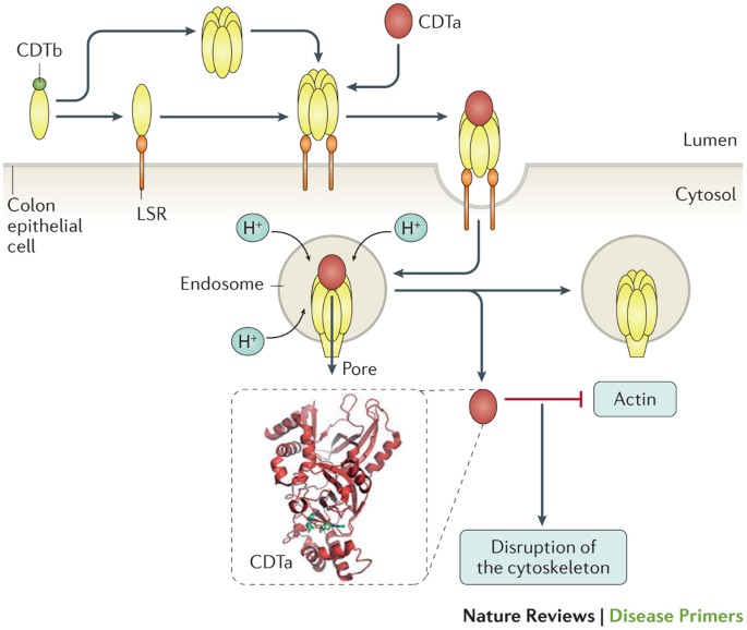
In some C.diff isolates, a binary toxin called C.diff transferase, has been isolated and found to cause a complete destruction of the actin cytoskeleton and therefore cell death. This toxin binds to the lipolysis-stimulated lipoprotein receptor in the gut. Here, it undergoes proteolytic activation and enters the cells via endocytosis. As the endosome matures, the V-ATPase decreases cellular pH and allows for the translocation of the toxin into the cytosol. The toxin then ribosylates actin at Arg177. This inhibits G-actin polymerization, therefore favoring F-actin depolymerization and causing actin cytoskeleton destruction. (Smits, Fig 7 to the left {above on mobile}, 2016)

Binding site
Clostridium difficile TcdA is a large toxin that binds carbohydrates on intestinal epithelial cells. ChimeraX cocrystal structure figure (above) of Clostridium difficile Toxin A fragment bound to two molecules of carbohydrate epitope aGal(1,3)bGal(1,4)bGlcNAc in an extended conformation. Toxin A Chain A is orange red. Chain B is hot pink. GAL, beta-D-galactopyranose is yellow. GLA, alpha-D-galactopyranose is dark blue. NAG, 2-acetamido-2-deoxy-beta-D-glucopyranose is light sea green. The Combined Repetitive Oligopeptides domain of C.diff toxins allows them to bind to carbohydrate receptors on host cells, aiding in their endocytosis. Residues forming key contacts with the trisaccharides are conserved in all seven putative binding sites in TcdA, suggesting a mode of multivalent binding that may be exploited for the rational design of novel therapeutics. (Greco, 2006)
Another failed attempt at fighting back
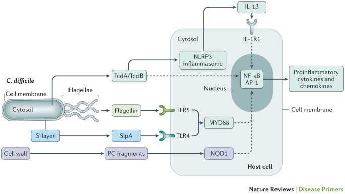
C.diff toxins A and B are recognized by the NLRP3 inflammasome, which directly leads to the production of Interleukin-1 beta. This proinflammatory cytokine plays a crucial role in the body’s immune response. The toxins also indirectly activate NF-kB and AP-1, which release more proinflammatory cytokines and chemokines. Fragments of the C.diff cell wall, surface layer protein A, and flagella also indirectly activate NF-κB, a key transcription factor involved in a host’s immune response. Cell wall peptidoglycan fragments are recognized by the nucleotide binding oligomerization domain containing protein 1 while flagellin and SlpA are recognized by toll-like receptors and activate the myeloid differentiation primary response 88. NOD1 and MYD88 then indirectly activate NF-kB and AP-1, leading to upregulation of proinflammatory cytokines secreted by lymphocytes, macrophages, and dendritic cells, including TNF-α, IL-6, and IL-8. (Kelly, 1994) (Smits, Fig 3 to the left {above on mobile}, 2016) The inflammatory response contributes to the colonic injury, with the release of inflammatory mediators from immune cells exacerbating tissue damage and promoting necrosis. This additionally causes damage to highly sensitive (due to their skinny nature) enteric neurons. These neurons are already impaired by the loss of electrolytes due to malabsorption, the production of ROS, and are very skinny, causing them to be one of the first parts of the body to accumulate damage. This affects intestinal motility, potentially leading to gut stasis and further exacerbating symptoms like constipation or dysmotility. (Kelly, 2008)
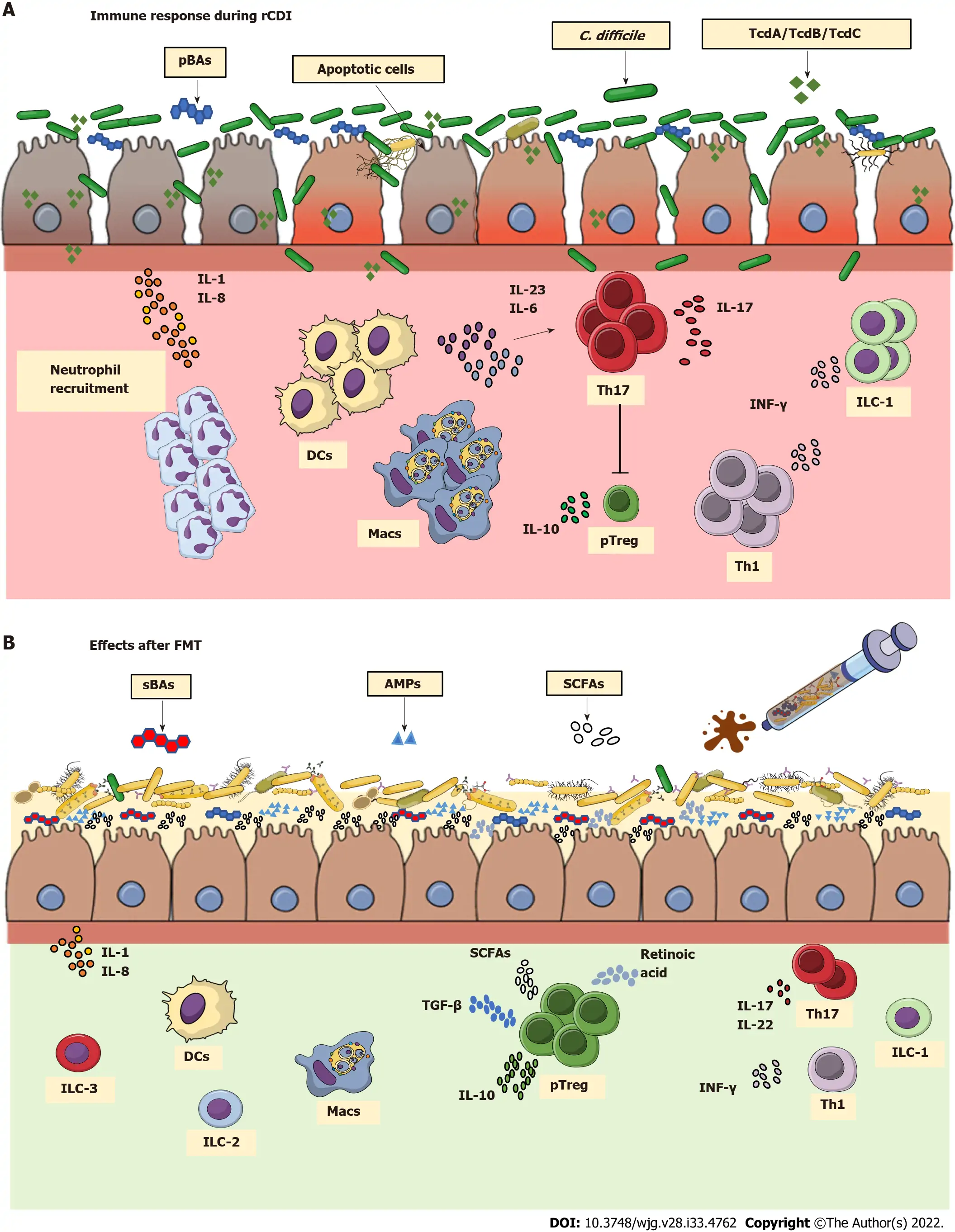
During recurrent C. difficile infection, the depletion of commensal microbiota results in higher levels of primary bile acids (BAs). These molecules are known to trigger the C. diff vegetative state and its expansion, resulting in production of toxins, which cause apoptosis of enterocytes and release of interleukin (IL)-1 and IL-8. Consequently, there is extensive recruitment of APCs in an attempt to cope with the infection. These cells produce pro-inflammatory cytokines which induce Th17 and Th1 cell differentiation. This inflammatory state promotes tissue damage. The therapeutic effects of fecal microbiota transplant, a therapeutic treatment for recurrent C.diff infection, involve the reestablishment of a wide variety of commensal microorganisms that directly/indirectly antagonize C. diff. Commensal strains that produce secondary BAs and short chain fatty acids like butyrate are re-established. The production of antimicrobial peptides by epithelial cells is also restored, allowing for the reconstitution of cell barrier integrity and deactivation of the innate immune response. (Soveral, 2022, Figure 2, to the left {above on mobile})
Small molecule dysfunction
Mitochondrial dysfunction activates ER stress proteins, Bax/Bak, leading to cytochrome C release, then caspase activation which causes apoptosis, ulceration, and diarrhea. (Voth, 2005) Mitochondrial damage also reduces ATP production as the electron transport chain function and oxidative phosphorylation is impaired. This limits the energy available for membrane repair, immune response, and epithelial barrier maintenance, causing necrosis in severe cases. This causes a disruption of AMPK signaling that affects cell survival and repair, worsening colonic injury. Fully inhibited AMPK activation can prevent energy homeostasis. (Zhao, 2016) Mitochondrial damage also increases reactive oxygen species (ROS), which at high levels, promote necrosis, protein oxidation, and lipid peroxidation. This causes oxidative damage to DNA, lipids, and proteins, therefore causing mutations, apoptosis, and colonic damage. (Fiorentini, 1998) Additionally, butyrate, a major short chain fatty acid, is reduced during CDI due to dysbiosis. Butyrate is essential for colonocyte energy, so its depletion contributes to mucosal atrophy and impaired gut repair. (Arcay, 2024)
references
- Madan, Rajat, and William A. Petri. “Immune Responses to Clostridium Difficile Infection.” Trends in Molecular Medicine 18, no. 11 (November 2012): 658–66. https://doi.org/10.1016/j.molmed.2012.09.005.
- Jank, Thomas, Torsten Giesemann, and Klaus Aktories. “Rho-Glucosylating Clostridium Difficile Toxins A and B: New Insights into Structure and Function.” Glycobiology 17, no. 4 (April 2007): 15R-22R. https://doi.org/10.1093/glycob/cwm004.
- Kelly, C. P., S. Becker, J. K. Linevsky, M. A. Joshi, J. C. O’Keane, B. F. Dickey, J. T. LaMont, and C. Pothoulakis. “Neutrophil Recruitment in Clostridium Difficile Toxin A Enteritis in the Rabbit.” The Journal of Clinical Investigation 93, no. 3 (March 1994): 1257–65. https://doi.org/10.1172/JCI117080.
- Kelly, Ciarán P., and J. Thomas LaMont. “Clostridium Difficile–More Difficult than Ever.” The New England Journal of Medicine 359, no. 18 (October 30, 2008): 1932–40. https://doi.org/10.1056/NEJMra0707500.
- Voth, Daniel E., and Jimmy D. Ballard. “Clostridium Difficile Toxins: Mechanism of Action and Role in Disease.” Clinical Microbiology Reviews 18, no. 2 (April 2005): 247–63. https://doi.org/10.1128/CMR.18.2.247-263.2005.
- Fiorentini, C., A. Fabbri, L. Falzano, A. Fattorossi, P. Matarrese, R. Rivabene, and G. Donelli. “Clostridium Difficile Toxin B Induces Apoptosis in Intestinal Cultured Cells.” Infection and Immunity 66, no. 6 (June 1998): 2660–65. https://doi.org/10.1128/IAI.66.6.2660-2665.1998.
- Nusrat, A., C. von Eichel-Streiber, J. R. Turner, P. Verkade, J. L. Madara, and C. A. Parkos. “Clostridium Difficile Toxins Disrupt Epithelial Barrier Function by Altering Membrane Microdomain Localization of Tight Junction Proteins.” Infection and Immunity 69, no. 3 (March 2001): 1329–36. https://doi.org/10.1128/IAI.69.3.1329-1336.2001.
- Zhao, Baozhong, Lei Qiang, Joy Joseph, Balaraman Kalyanaraman, Benoit Viollet, and Yu-Ying He. “Mitochondrial Dysfunction Activates the AMPK Signaling and Autophagy to Promote Cell Survival.” Genes & Diseases 3, no. 1 (March 1, 2016): 82–87. https://doi.org/10.1016/j.gendis.2015.12.002.
- Arcay, Ricardo, Maria Barceló-Nicolau, Loreto Suárez, Luisa Martín, Rebeca Reigada, Marcus Höring, Gerhard Liebisch, et al. “Gut Microbiome and Plasma Lipidome Analysis Reveals a Specific Impact of Clostridioides Difficile Infection on Intestinal Bacterial Communities and Sterol Metabolism.” mBio 15, no. 10 (n.d.): e01347-24. https://doi.org/10.1128/mbio.01347-24.
- Bank, RCSB Protein Data. “RCSB PDB – 2BVL: Crystal Structure of the Catalytic Domain of Toxin B from Clostridium Difficile in Complex with UDP, Glc and Manganese Ion.” Accessed March 31, 2025. https://www.rcsb.org/structure/2BVL.
- Smits, Wiep Klaas, Dena Lyras, D. Borden Lacy, Mark H. Wilcox, and Ed J. Kuijper. “Clostridium Difficile Infection.” Nature Reviews. Disease Primers 2 (April 7, 2016): 16020. https://doi.org/10.1038/nrdp.2016.20.
- Greco, Antonio, Jason G. S. Ho, Shuang-Jun Lin, Monica M. Palcic, Maja Rupnik, and Kenneth K.-S. Ng. “Carbohydrate Recognition by Clostridium Difficile Toxin A.” Nature Structural & Molecular Biology 13, no. 5 (May 2006): 460–61. https://doi.org/10.1038/nsmb1084.
- Reinert, Dirk J., Thomas Jank, Klaus Aktories, and Georg E. Schulz. “Structural Basis for the Function of Clostridium Difficile Toxin B.” Journal of Molecular Biology 351, no. 5 (September 2, 2005): 973–81. https://doi.org/10.1016/j.jmb.2005.06.071.